Session highlights discoveries in myocardial regeneration
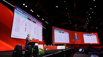
Potential treatment strategies to repair the heart are emerging through clinical studies, said presenters in Monday’s “New Frontiers in Myocardial Regeneration” session.
The session featured advances in various myocardial regeneration approaches and possible mechanisms of regeneration by applying animal models to human biology and novel strategies.
“Bringing a drug to market is expensive because the animal model and simple human cell culture are not a good predictor of human physiology,” said Gordana Vanjak-Novakovic, PhD, the University and Miakti Foundation professor of biomedical engineering and medical sciences laboratory for stem cells and tissue engineering at Columbia University in New York City.
Dr. Vanjak-Novakovic discussed how a novel research platform for engineering human myocardium, “organs on a chip,” involves bioengineering and maturing tissue on physical platforms, designed to emulate human physiology, can provide more relevant results to actual humans.
“We design the environments, and cells do the engineering,” she said.
Using a Google map as an analogy, Li Qian, PhD, described how she and her team at the McAllister Heart Institute at the University of North Carolina at Chapel Hill Direct are using single cell “transcriptonomics” and mathematical modeling to reconstruct the molecular roadmap of cardiac reprogramming to ultimately convert cardiac fibroblasts into cardiomyocyte-like cells.
“We hope to treat cardiac fibroblasts to create personalized treatments for myocardial infarction,” she said.
In her four vignettes about cardiac repair, Nadia Rosenthal, PhD, scientific director of The Jackson Laboratory in Bar Harbor, Maine, also presented insight into the complexity of myocardial regeneration.
“Cardiac cellular composition is dynamic, sexually dimorphic and altered by stress/damage signals,” she said.
Moreover, cardiac fibroblasts retain a regional code of morphogenetic gene expression, possibly for tissue repair. Her research reveals that immune interstitial signaling circuits provide important novel entry points for improving repair.
“Cardiac repair as a genetically tractable trait offers prospects for mapping gene networks and therapeutic targets,” she said.
Mauro Giacca, MD, PhD, professor of cardiovascular sciences in the School of Cardiovascular Medicine & Sciences at King’s College in London, presented his research on lipid-based nanoparticle (LNP) microRNA (miRNA) networks.
“Throughout life, we continue to lose cardiomyocytes,” he said. “All the drugs we have only target the capacity of the residual myocytes to improve their function. New treatments are needed to generate new cardiomyocytes. We have two options: Implant newer cells from the outside or try to convince surviving cardiomyocytes to proliferate.”
Dr. Giacca described how LNPmiRNA therapy delivered via intramyocardial injection may be able to transform non-cardiomyocytes into proliferating cells.
“The idea of using LNP-miRNA for cardiac regeneration is possible,” he said. “Even if you gain 10% to 20% ejection fraction, that can prevent heart failure.”
Jeff D. Molkentin, PhD, director of the division of Molecular Cardiovascular Biology at Cincinnati Children’s in Ohio, concluded the session with an overview of his research on myocardial cell therapy for myocardial cell rejuvenation: Injecting bone marrow mononuclear cells into the heart induces endothelial cell activity and new vascular genesis.
His research suggests that the acute sterile immune response from the injection therapy induces angiogenesis in the heart at injection sites, reconditions the extracellular membrane and reduces border zone fibrosis expansion.
“It’s all just wound-healing biology,” Dr. Molkentin said.
