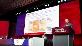
Late-breaking trials showcase AI’s potential as a cardiovascular disease screening and diagnostic tool

Trial results presented in Monday’s Late-Breaking Science session provided insight into the potential role for artificial intelligence to guide cardiac care. Their results found:
- An AI-enabled smartwatch has potential for identifying left ventricular systolic dysfunction.
- AI assistance for transthoracic echocardiogram shows promise as a transthoracic echocardiogram tool.
- AI assistance can generate a comprehensive and accurate TTE interpretation.
AI-enabled smartwatch shows promise as an ECG screening tool
AI applied to a consumer smartwatch electrocardiogram (ECG) could offer a promising new approach to screening for left ventricular systolic dysfunction (LVSD) or reduced heart pumping function, according to findings from the WATCH-LV trial. The study, “Consistency of Smartwatch Artificial Intelligence-Enabled Electrocardiogram for Left Ventricular Systolic Dysfunction Across Manufacturers and Repeated Home Recordings,” evaluated the technology’s performance across two smartwatch brands.
The prospective, single-center study included a hospital-based cohort of 2,211 participants undergoing clinically indicated echocardiography who recorded ECGs using both an Apple Watch and a Samsung Galaxy Watch under medical supervision. A separate home-monitoring cohort included 146 participants, 49 of whom had LVSD, defined as a left ventricular ejection fraction of ≤40% on echocardiography. The study examined 2,984 recordings obtained during the first seven days, comparing individual recordings, averaged scores and rules requiring repeated positive results before triggering an alert.
 Hak Seung Lee, MD
The home-monitoring findings highlighted the importance of how repeated results are interpreted. Among participants without LVSD who recorded on at least five days, approximately 26% had at least one positive recording day during the week. Requiring positive results on at least two of three successive recording days reduced this proportion to approximately 13%.
LVSD was present in approximately 81% of participants whose recordings were consistently positive, compared with approximately 6% of those whose recordings were consistently negative. These proportions apply to this selected study population and should not be interpreted as an individual consumer’s probability of disease, Lee said. Averaging repeated recordings produced numerically higher discrimination than a single recording, but the difference was not statistically significant.
Overall, “smartwatch ECG recording and AI-ECG research have reached a point at which we can begin addressing practical questions about use beyond the clinic,” Lee said. “Demonstrating that an algorithm performs well on an individual ECG is an important first step.” He noted that larger, more diverse prospective studies are needed to confirm these findings. If confirmed, smartwatch AI-ECG could serve as a screening tool that complements, rather than replaces, clinical assessment and echocardiography.
AI-assisted interpretation of transthoracic echocardiograms was noninferior to sonographer and cardiologist interpretations
AI could significantly reduce the time cardiologists spend interpreting echocardiograms without compromising accuracy, according to the findings from the trial “Randomized Controlled Trial of AI-Assisted Echocardiography Interpretation: AI Echo Insight.”
This investigator-initiated, blinded, randomized, noninferiority trial utilized 1,200 transthoracic echocardiograms (TTEs) acquired consecutively in March 2025 across the 21 medical centers of Kaiser Permanente Northern California. Of the 1,200 TTEs, 200 TTEs with at least moderate valvular pathology were included to enrich interpretation complexity. For each of the 1,200 TTE examinations in the study cohort, three preliminary reports were generated: one by a cardiologist, one by a sonographer and one by the EchoPrime AI model developed using data outside of Kaiser Permanente. Subsequently, 12 adjudicating cardiologists blinded to the source of the preliminary interpretations independently over-read the reports. The accuracy of the AI-assisted arm was compared with a pooled comparator arm of cardiologists and sonographers in a noninferiority analysis with a prespecified margin of five percentage points. The primary endpoint was substantial change between preliminary and over-read interpretations, comparing the AI-assisted workflow to the pooled cardiologist-assisted and sonographer-assisted workflows.
The over-reading cardiologists, asked to guess the source of each preliminary report they reviewed, could not determine whether it was performed by AI or another clinician, confirming sufficient blinding. The accuracy of the AI-generated preliminary interpretations was noninferior to that of the pooled comparator of sonographer- and cardiologist-generated preliminary interpretations. With AI assistance, cardiologists spent 4.6 minutes interpreting a TTE, compared with roughly 8.5 minutes per TTE without any assistance, a time savings of approximately 46% per interpretation. The AI-assisted finalized reports were also more consistent with the historical reports in the medical record compared to those generated with sonographer and cardiologist assistance, indicating that AI could improve the precision of reporting.  Keane Lee, MD, MS
Overall, AI preliminary interpretation was noninferior to pooled sonographer and cardiologist preliminary interpretation. “We demonstrated that AI can generate a comprehensive and accurate interpretation of a full TTE examination. This technology has the capability to significantly reduce cardiologist and sonographer time usage, while improving the precision of interpretations,” said Keane Lee, MD, MS, a cardiologist at Kaiser Permanente Santa Clara and study principal investigator. He noted that the Kaiser Permanente Northern California health system is implementing EchoPrime AI assistive technology for TTE interpretation. “EchoPrime helps us work more efficiently, giving time back to our cardiologists and sonographers while also improving the precision of our interpretations. It’s clearly a win on multiple fronts,” he said.
AI-enabled tele-ECG highlights potential role in SHD screening
Artificial intelligence-enabled electrocardiography (AI-ECG) outperformed conventional assessment of structural heart disease (SHD), according to “AI-Enabled Tele-ECG Identifies Structural Heart Disease and Prioritizes Echocardiography in Underserved Populations: The PROVAR+ External Validation Study.”
The prospective observational study evaluated digital 12-lead tele-ECGs acquired within the Telehealth Network of Minas Gerais, and comprehensive transthoracic echocardiography (TTE) of 3,779 adults receiving care within PROVAR+, a Brazilian cardiovascular screening initiative integrating primary care, telemedicine, portable imaging and specialist interpretation. Tests were conducted on the same day whenever possible over 11 days in 2025 during cardiovascular screening efforts in two Brazilian cities.
Participants were identified through public TTE waiting lists following clinical referral or tele-ECG and handheld echocardiography screening because of major ECG abnormalities or abnormal preliminary imaging, including a predefined sample without major ECG abnormalities. Median age was 66; 56.7% were women. Pre-existing conditions included hypertension (73.1%), diabetes (34.6%), heart failure (19.9%), coronary artery disease (16.5%) and stroke (7.9%).
Cardiologists interpreted the digital tele-ECGs; ECG waveforms were also processed using a previously developed and validated SHD algorithm assessed through a cloud-based application programming interface (API). The model generated a continuous probability of SHD. Its binary performance was evaluated using a prespecified threshold of 0.204646, defined in preliminary multinational studies. AI inference was applied post hoc and did not determine patient referrals.
The reference standard was comprehensive TTE interpreted by board-certified cardiologists blinded to AI-ECG results, with additional adjudication when necessary. Clinically significant SHD included ventricular dysfunction or hypertrophy, regional wall-motion abnormalities, significant valvular disease, congenital abnormalities and pericardial disease. Three prediction strategies were compared: AI-ECG probability alone; a conventional model combining demographic and clinical variables with conventional major ECG abnormalities; and a combined model incorporating AI-ECG probability, clinical variables and conventional ECG interpretation.
AI-ECG identified 87.3% of patients with significant SHD and performed better than conventional ECG interpretation combined with clinical information (AUROC 0.80, 95% CI: [0.78, 0.82]). AI-ECG alone differentiated SHD better than traditional ECG/clinical assessment plus ECG interpretation, generating fewer unnecessary echocardiography referrals. Bruno Ramos Nascimento, MD, MSc, PhD, FACC, FESC
“Most importantly, our simulations suggested that AI-guided prioritization could reduce the estimated time needed to diagnose half of affected patients from 407 to 172 days, a 58% reduction, consistent across genders and age groups,” said Bruno Ramos Nascimento, MD, MSc, PhD, FACC, FESC, associate professor of interventional cardiology at the Federal University of Minas Gerais in Brazil, the principal investigator. “Our study shows the potential to use existing imaging resources better without needing more echocardiography capacity, and how to optimize health resources.”
Nascimento noted that this technology could help prioritize patients for heart imaging, particularly in healthcare systems facing long waiting lists and limited resources. Further pragmatic implementation studies are essential to determine whether these strategies can translate into earlier diagnoses, more equitable access to care and, ultimately, better health outcomes, he said. The study was published in The American Journal of Cardiology following the presentation.